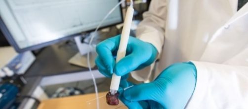
Researchers at the University of Texas have designed a real-time diagnostic tool that can detect cancer cells within seconds, states a new study published in the journal Science Translational Medicine on Wednesday.
About the ‘cancer pen’
Known as the MasSpec pen, the Handheld Device can analyse a tissue’s molecular composition in ten seconds, once the doctor places the pen on a patient’s tissue. The MasSpec pen uses tiny water droplets to read human tissue samples and identify it as either normal or cancerous.
The cells in our body produce a range of tiny molecules.
Cancer develops a unique set of these human cells that can be used for pattern identification, explains Livia Schiavinato Eberlin, study author and Assistant Professor of Chemistry at UT Austin, in a TIME report.
Researchers tested the prototype on more than 250 samples of human tissue samples from thyroid, lung, ovary and breast cancer tumours and compared them to healthy tissue samples. The instrument was 96% accurate in distinguishing between healthy and cancerous tissue, noted the Scientists.
Also, the device is able to identify different subtypes of lung and thyroid cancer as well. Scientists will now focus on making its analysis more specified for other types of cancer too. The team is hopeful that the pen-sized prototype could be used in clinical trials in early 2018.
"I see this as a very interesting type of technology that will probably have a place in surgical oncology," Dr John Rudan, Head of Surgery at Queen's University in Kingston, Ontario, tells CBC News. Rudan was not involved in the study.
The team, working with funding from the National Cancer Institute and the Cancer Prevention Research Institute of Texas, has filed patents for the technology.
A look at similar experimental technologies
The MasSpec is not the only latest technology that’s aimed at improving cancer surgery.
In February, the University of Michigan Medical School and Harvard University developed a technique called SRS microscopy. The technology uses laser beams to help surgeons decide how much tissue needs to be removed by swiftly analysing brain cancers.
“The differing chemistry of a cancerous cell and normal brain tissue means the laser help surgeons find the outside edge of a tumour,” says a BBC report.
Last year, researchers at Duke University Medical Center, Massachusetts General Hospital and Massachusetts Institute of Technology developed a probe that could detect and light up residual cancer cells, making it easier for surgeons to spot them. The new imaging technique could make cancer surgery more effective in future. Also, it could help doctors “to target radiation therapy to just the right parts of a tumour to kill remaining cancer cells,” says TIME.
In 2013, researchers at the Imperial College of London modified a surgical knife that could cut down the risk of tumour recurrence. Dubbed as the iKnife, the instrument can be used to accurately identify cancerous tissue by detecting the subtle differences between the smoke emanating from a cancerous and healthy tissue.


